What Is An MRI Arthrogram?
An MRI arthrogram in our Gold Coast clinic involves a CT guided injection of MRI contrast dye (Gadolinium) into a joint to enhance the diagnostic quality of the MRI scan by allowing more detailed visualisation of the fine structures within the joint, such as cartilage and ligaments.
Arthrograms are usually performed under CT guidance before an MRI scan, most commonly for the shoulder or hip. The Gold Coast based radiologists at Panorama Radiology Specialists have undergone advanced subspecialist training in interventional and musculoskeletal radiology, and have extensive clinical experience in performing and reading MRI arthrograms. Typically the procedure is performed under CT guidance and local anaesthesia, and is usually quick, safe and involves minimal discomfort. The contrast dye passes from the body within a few hours.
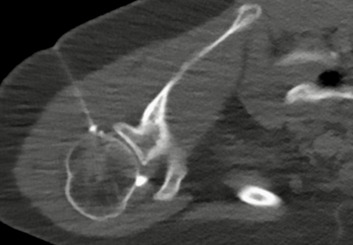
An MRI arthrogram allows the radiologist and your orthopaedic surgeon or other treating clinician to accurately assess injuries to the fine structures of the joint, including labral and ligament tears and cartilage injuries, which may not always be visible on a standard MRI scan.
A common reason for performing MRI arthrography is prior shoulder dislocation or recurrent shoulder instability, to allow accurate detection of labral tears that may require surgical management or hip pain for suspected labral tears or femoro-acetabular impingement.
An MRI arthrogram allows accurate diagnosis of the nature and extent of shoulder and hip injuries, which assists shoulder surgeons with operative planning, and helps sports medicine practitioners, physiotherapists, WorkCover practitioners and other clinicians to obtain an accurate diagnosis to accurately inform management and rehabilitation for a variety of shoulder and hip conditions, including – acute and chronic injuries, instability, clicking hips or shoulders, rotator cuff tears, frozen shoulders and chronic impingement syndromes.